|
What is Patellar Luxation?
MEDIALLY LUXATING PATELLA (MLP)
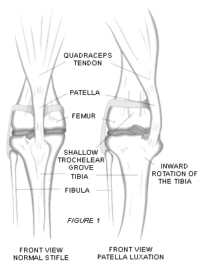
Medial patella luxation is typically seen as a squat gait and toed-in appearance related to the severity of the condition.
The patella may pass over the midline axis of the stifle joint in the lateral projection such that the quadriceps becomes
a flexor rather than an extensor of the limb; these dogs are unable to extend their legs while walking. The crouched gait
of the severely affected animal is almost pathognomonic. Occasionally, in animals not affected severely, little is seen on
gaiting the animal. Occasional hopping or skipping of a beat of a stride is an indication to check the stifle joints. Lateral
patellar luxation usually results in a very straight-legged appearance, since the dog uses a shuffling gait with a shorter
stride behind. Both conditions may be associated with cruciate rupture and may then present with only an indication of a lame
leg. Hyperextension of the hock joints is sometimes seen in these straight-legged dogs and is evidenced in standing and walking.
Medially Luxating Patella
(also described as slipping patella or knee cap)
by Dr Steven Metcalf BSc BVMS (Hons) MSc MACVSc
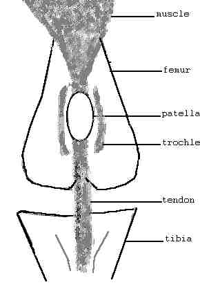
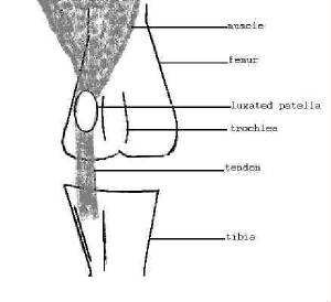
Medially luxating patella (MLP) refers to the tendency for the kneecap to slip out of its normal groove and to become trapped
on the inside of the knee or stifle joint. It is a common cause of hind limb lameness in small breed dogs but will occur in
dogs of all ages, sizes and breeds.
During growth and development the quadriceps muscles in front of the thigh tends to pull the kneecap to the inside of the
leg rather than pulling the kneecap up and down the trochlear groove. Over time the inside ridge of the trochlear groove erodes
and the groove fails to deepen because of the absence of normal wear.
Lameness occurs because the kneecap does not have a normal groove to slide in. With time joint restriction and osteoarthritis
may develop.
Severity of MLP is determined by palpation of the knee joint and is classified from 0 (normal) to 4 (severely affected) as
follows;
Grade 0 Normal
Grade 1 the patella can be pushed out of the groove but spends most of time in the groove
Grade 2 the patella approximately half the time in the groove and half the time out of the groove. It can easily be manipulated
in or out but has no tendency to stay in either position.
Grade 3 the patella spends almost all the time outside the groove and with pressure can be pushed back into the groove.
Grade 4 the patella spends all the time outside the groove and even with pressure the patella cannot be pushed back into
the groove.
© Dr Steven Metcalf BSc BVMS (Hons) MSc MACVSc
Applecross Veterinary Hospital
Perth, Western Australia
Patellar Luxation Treatment
TREATMENT
Grade 1 and early grade 2 MLP may only require medical treatment for pain and minor swelling within the knee joint. Mid-stage
grade 2, grade 3 and grade 4 are best corrected with surgery. The three common surgical procedures are described as follows;
Tibial Crest Transposition (TCT). The patella tendon attaches to the top of the tibia. This area of bone is surgically
separated from the shaft of the tibia and repositioned on the outside of the tibia. The bone is secured by pins and wires
or a surgical screw. Recovery time to normal walking is 6-8 weeks although most dogs are weight bearing within 1 to 2 days.
Wedge Recession Trochleoplasty (WRT). The groove in the femur where the patella lies is surgically deepened by cutting
out a wedge shaped section.
Lateral ligament placement. This involves placing nylon bands through the patella and its tendon and anchoring them to
the outside of the knee joint.
Any, a combination of, or all of the above techniques may be used to repair MLP. This decision is made during surgery
and depends on the size of the dog, degree of luxation, conformation of the knee joint and the presence of osteoarthritis.
The prognosis following surgery is good to excellent in at least 95% of cases. Those cases that have a poorer prognosis
are those with poor knee conformation, grade 3 and 4 luxations and those knees with osteoarthritis.
© Dr Steven Metcalf BSc BVMS (Hons) MSc MACVSc
Applecross Veterinary Hospital
Perth, Western Australia
Patellar Luxation Links:
Click Here for Vet Surgery Patella Page
Click here for Textbook of Small Animal Orthopedics Patellar luxation (has some surgical techniques and everything!
Click here for Working Dogs Patellar Luxation. Has GREAT information and GREAT illustrations of patellar luxation!
Merck Veterinary Manual Patellar Luxation
|